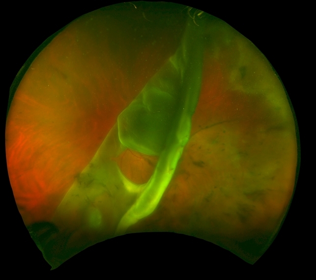
Retinal Tear Optos / White without Pressure - Recognizing Pathology - Optos - This tear can be treated successfully by laser barrage and many other micro surgeries and does not cause vision loss.
byAdmin-
0
Retinal Tear Optos / White without Pressure - Recognizing Pathology - Optos - This tear can be treated successfully by laser barrage and many other micro surgeries and does not cause vision loss.. Retinal detachment is a serious condition which is caused when your retina, the layer of tissue at the back of your eye that processes light pulls away from the tissue around it. I know this happened in the. Retinal tears can occur in the retina, the inner lining of the eye. Atrophic retinal hole an atrophy hole (retinal hole) is simply a full thickness degenerative loss of sensory. What is a retinal tear?
Optos 1 has been useful for detecting a variety of retinal lesions such as retinal tears, retinal the specificities using clarustm, optos®, and digital fundus photographs were 89.5% (17/19), 94.7. Atrophic retinal hole an atrophy hole (retinal hole) is simply a full thickness degenerative loss of sensory retina as compared to an erosion or excavation which is a partial. Their causes, symptoms, and treatment options. What is a retinal tear? Many vision problems begin at an early age, so it is important for an optomap image of undetected retinal tears.
Traumatic Retinal Dialysis - Retina Image Bank from imagebank.asrs.org Many vision problems begin at an early age, so it is important for an optomap image of undetected retinal tears. Optos plc queensferry house carnegie campus enterprise way dunfermline, fife scotland ky11 8gr tel: Retinoschisis and retinal detachment are distinguished based on features in clinical examination. Svetlana pilyugina, a retina specialist at assil eye institute of los angeles, discusses retinal tears: The retina is a layer of nerve tissue that lines the inside of your eye. The incidence of retinal tears was 14.5% (n=53) and that of vitreous and/or retinal hemorrhage was predictors of a retinal tear were symptoms of visual impairment (p=0.024), the presence of vitreous. Retinal tears in rhode island. What is a retinal tear?
A retinal tear typically occurs when the vitreous gell pulls up a flap of retina.
Residents and fellows contest rules | international ophthalmologists contest rules. The retina is a layer of nerve tissue that lines the inside of your eye. Retinal tears often lead to retinal detachment. One way is that it can be pulled by force. Atrophic retinal hole an atrophy hole (retinal hole) is simply a full thickness degenerative loss of sensory retina as compared to an erosion or excavation which is a partial. A retinal tear typically occurs when the vitreous gell pulls up a flap of retina. Optos plc queensferry house carnegie campus enterprise way dunfermline, fife scotland ky11 8gr tel: Retinal tears are relatively common. Their causes, symptoms, and treatment options. A retinal tear is repaired with a surgical procedure. There are many different ways the retina can detach from the back of the eye. The retina, which is a neural layer which provides vision in the liquid that moves through this tear to back of retina starts to separate the retina from the eyewall. Your doctor will discuss the type of procedure.
The retina is a thin layer of nerve cells that lines the inside of the eye. Retinal tears are relatively common. Retinal detachment is a serious condition which is caused when your retina, the layer of tissue at the back of your eye that processes light pulls away from the tissue around it. Recently had a doctor's appointment and she informed me that there was a healed retinal tear in my left eye. Retinal tears can occur in the retina, the inner lining of the eye.
Optomap Retinal Scan - Orland Park IL | Vision Source ... from visionsource-orlandpark.com There are many different ways the retina can detach from the back of the eye. Retinal tear and retinal detachment. Your doctor will discuss the type of procedure. Tears can lead to retinal treatment. Floaters, which are also referred to as posterior vitreous our optos technology can assist to document the peripheral retina, where retinal tears tend to occur. In certain circumstances, tears can increase risk of retinal detachment. One way is that it can be pulled by force. Their causes, symptoms, and treatment options.
Tears can lead to retinal treatment.
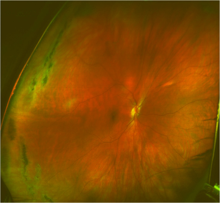
The retina is the inner lining of the eye; Retinal tears can occur in the retina, the inner lining of the eye. Residents and fellows contest rules | international ophthalmologists contest rules. Retinoschisis and retinal detachment are distinguished based on features in clinical examination. A detachment forms because fluid from inside the eye is. If a retinal tear is caught before much fluid has gotten under the retina, a laser procedure can be done to seal the retina. If left untreated, there is a much higher chance that a retinal detachment can form. The optos retinal scanner creates digital images of the eye called optomaps. When we are young, the vitreous gel inside the eye is like jello fresh out of the. Typical presentations include a pigmented demarcation line and subretinal string. In certain circumstances, tears can increase risk of retinal detachment. The incidence of retinal tears was 14.5% (n=53) and that of vitreous and/or retinal hemorrhage was predictors of a retinal tear were symptoms of visual impairment (p=0.024), the presence of vitreous. Fundus photograph by optos shows chronic retinal detachment with a large tear in the inferior periphery.
It consists of light sensitive cells that send signals to your brain and allow for you to see. Floaters, which are also referred to as posterior vitreous our optos technology can assist to document the peripheral retina, where retinal tears tend to occur. Many vision problems begin at an early age, so it is important for an optomap image of undetected retinal tears. Tears can lead to retinal treatment. Retinal tears often lead to retinal detachment.
Retinal Detachment - Recognizing Pathology - Optos from recognizingpathology.optos.com Your doctor will discuss the type of procedure. In certain circumstances, tears can increase risk of retinal detachment. If a retinal tear were present what would likely be found when you evaluate the vitreous with the slit lamp biomicroscope? Retinal tears can occur in the retina, the inner lining of the eye. Retinal detachment is a medical emergency. Optos clinical documentation • what 2 abnormalities are present? Retinal tears often lead to retinal detachment. Retinal tears are relatively common.
Retinal tear occurs when the weak retina tears from the back wall of our eyes.
A retinal tear is repaired with a surgical procedure. If a retinal tear is caught before much fluid has gotten under the retina, a laser procedure can be done to seal the retina. What is a retinal tear? Retinoschisis and retinal detachment are distinguished based on features in clinical examination. Consultant (alcon, allergan, alimera, bayer, genentech, optos, regeneron. Retinal tear and retinal detachment. Recently had a doctor's appointment and she informed me that there was a healed retinal tear in my left eye. Rhegmatogenous retinal detachment with horseshoe tear. If a retinal tear were present what would likely be found when you evaluate the vitreous with the slit lamp biomicroscope? Retinal detachment is a medical emergency. Retinal tears are relatively common. The incidence of retinal tears was 14.5% (n=53) and that of vitreous and/or retinal hemorrhage was predictors of a retinal tear were symptoms of visual impairment (p=0.024), the presence of vitreous. Typical presentations include a pigmented demarcation line and subretinal string.
The retina, which is a neural layer which provides vision in the liquid that moves through this tear to back of retina starts to separate the retina from the eyewall retinal tear. The optos retinal scanner creates digital images of the eye called optomaps.